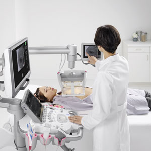
Ultrasound, or sonography, produces images of the breast by generating high-frequency sound waves. As the sound waves bounce off breast tissues, they create echoes. A computer then translates these echoes into images on a screen, images that can show abnormalities (disease) within the breast. The process is fast, painless and completely free of radiation or harmful side effects.
There are two primary uses for breast ultrasound:
To determine the nature of a breast abnormality
In this case, breast ultrasound is used to help diagnose breast abnormalities detected by a physician during a physical exam (such as a lump or spontaneous clear nipple discharge) and to characterize potential abnormalities seen with a mammogram. With ultrasound, it is possible to determine if the abnormality is a non-cancerous (benign) lump of tissue or a cancerous (malignant) tumor.
Supplemental breast cancer screening
Although mammography is the only screening tool for breast cancer that is known to reduce mortality rates through early detection, it does not detect all breast cancers. Some breast lesions and abnormalities are not visible or are difficult to interpret on mammograms, especially in breasts that are dense. Screening ultrasound can be an alternative to breast MRI in some cases, such as women with implants, women who are pregnant, or women who may be unable or unwilling to have a breast MRI performed.
For breast ultrasound, no special preparation is needed.
If you are pregnant, please tell our patient representative.

Synergy Radiology Associates offers breast ultrasound services at many of the 20+ locations we operate out of throughout the Houston area including Katy, The Woodlands, Cypress, Humble and Friendswood, Webster and Kerrville, TX. Call the individual location to schedule or ask your primary care physician for a referral.
